Heading 1
Heading 2
Heading 3
Heading 4
Heading 5
Heading 6
Lorem ipsum dolor sit amet, consectetur adipiscing elit, sed do eiusmod tempor incididunt ut labore et dolore magna aliqua. Ut enim ad minim veniam, quis nostrud exercitation ullamco laboris nisi ut aliquip ex ea commodo consequat. Duis aute irure dolor in reprehenderit in voluptate velit esse cillum dolore eu fugiat nulla pariatur.
Block quote
Ordered list
- Item 1
- Item 2
- Item 3
Unordered list
- Item A
- Item B
- Item C
Bold text
Emphasis
Superscript
Subscript
About This Simulation
Join doctors in revealing a pathogen that is causing a patient to be critically ill. Perform the Gram stain on a sample collected from the patient and use microscopy to identify the presence of bacteria to help guide the proper antibiotic treatment.
Learning Objectives
- Describe the structure of the Gram-positive and Gram-negative bacteria
- Appreciate theoretical and technical aspects of the Gram staining procedure
- Know the most commonly made mistakes in Gram staining
- Critically interpret the results of a Gram staining experiment using a light microscope
About This Simulation
Lab Techniques
- Light microscopy
- Preparation of bacterial smears
- The Gram stain technique
Related Standards
- No direct alignment
- No direct alignment
- B.1 Microbiology: organisms in industry
Learn More About This Simulation
Did you know that there are approximately 5 million-trillion-trillion bacteria in the world? Most of them are harmless, but some can induce disease in an affected host. In this simulation, you will help doctors identify bacteria in a cerebrospinal fluid sample from a patient suspected of suffering from bacterial meningitis.
Explore the bacterial cell wall
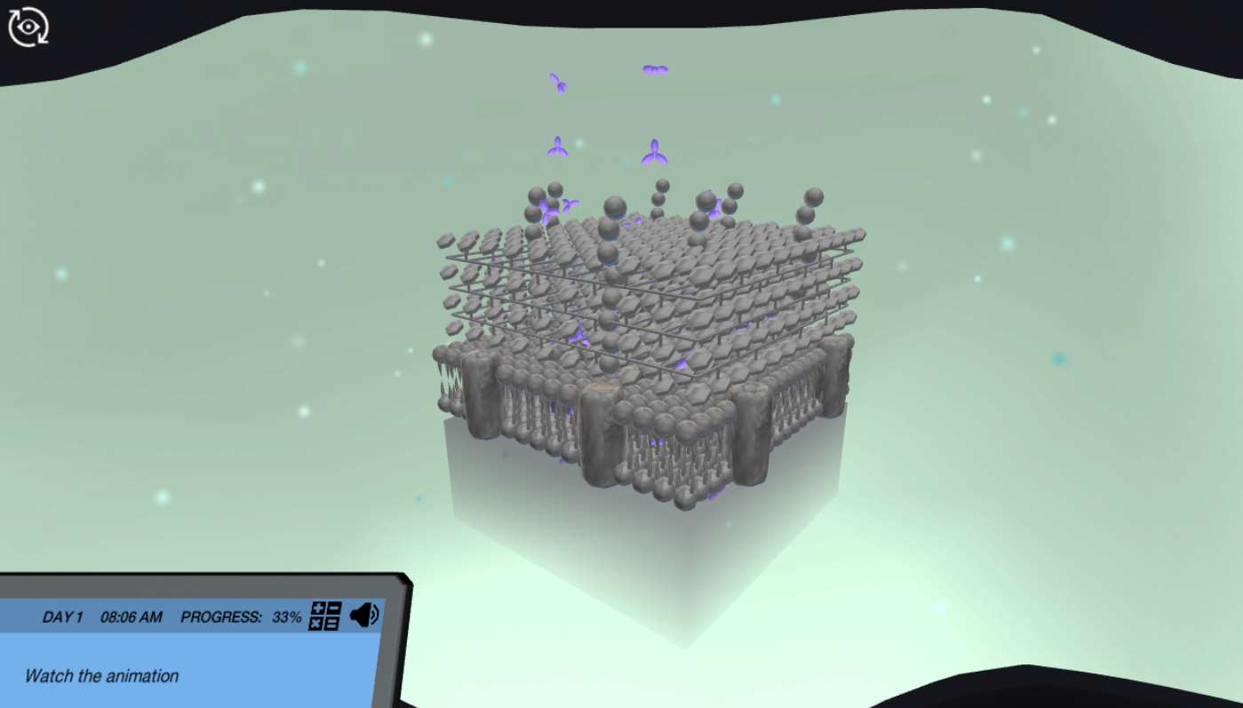
Compare and contrast the cell wall of Gram-positive and Gram-negative bacteria by building your very own bacterial 3D models on the hologram table. Enter the exploration pod to observe in an immersive animation how the four reagents of the Gram stain interact with structural components of the cell wall to color the bacteria.
Perform the Gram stain
When the patient's fluid sample arrives at the laboratory, equip yourself with protective gear to prepare a bacterial smear and heat fix it to a glass slide. You are now ready to perform the Gram stain in a safe virtual environment. Made a mistake? No worries, hit the big red button on the workbench to repeat the staining procedure until it becomes second nature.
Interpret your findings using a microscope
In the end, you will use a light microscope to interpret the results of your Gram stain. View the microscopic image on the computer screen, and apply immersion oil to increase magnification 1000x! Will you be able to identify the presence of any bacteria in the patient´s cerebrospinal fluid?
For Science Programs Providing a Learning Advantage
Boost STEM Pass Rates
Boost Learning with Fun
75% of students show high engagement and improved grades with Labster
Discover Simulations That Match Your Syllabus
Easily bolster your learning objectives with relevant, interactive content
Place Students in the Shoes of Real Scientists
Practice a lab procedure or visualize theory through narrative-driven scenarios


FAQs
Find answers to frequently asked questions.
Heading 1
Heading 2
Heading 3
Heading 4
Heading 5
Heading 6
Lorem ipsum dolor sit amet, consectetur adipiscing elit, sed do eiusmod tempor incididunt ut labore et dolore magna aliqua. Ut enim ad minim veniam, quis nostrud exercitation ullamco laboris nisi ut aliquip ex ea commodo consequat. Duis aute irure dolor in reprehenderit in voluptate velit esse cillum dolore eu fugiat nulla pariatur.
Block quote
Ordered list
- Item 1
- Item 2
- Item 3
Unordered list
- Item A
- Item B
- Item C
Bold text
Emphasis
Superscript
Subscript
A Labster virtual lab is an interactive, multimedia assignment that students access right from their computers. Many Labster virtual labs prepare students for success in college by introducing foundational knowledge using multimedia visualizations that make it easier to understand complex concepts. Other Labster virtual labs prepare learners for careers in STEM labs by giving them realistic practice on lab techniques and procedures.
Labster’s virtual lab simulations are created by scientists and designed to maximize engagement and interactivity. Unlike watching a video or reading a textbook, Labster virtual labs are interactive. To make progress, students must think critically and solve a real-world problem. We believe that learning by doing makes STEM stick.
Yes, Labster is compatible with all major LMS (Learning Management Systems) including Blackboard, Canvas, D2L, Moodle, and many others. Students can access Labster like any other assignment. If your institution does not choose an LMS integration, students will log into Labster’s Course Manager once they have an account created. Your institution will decide which is the best access method.
Labster is available for purchase by instructors, faculty, and administrators at educational institutions. You can purchase Labster by speaking with a Labster sales representative. Compare plans.
Labster supports a wide range of STEM courses at the high school, college, and university level across fields in biology, chemistry, physics, and health sciences. You can identify topics for your courses by searching our Content Catalog.


