Heading 1
Heading 2
Heading 3
Heading 4
Heading 5
Heading 6
Lorem ipsum dolor sit amet, consectetur adipiscing elit, sed do eiusmod tempor incididunt ut labore et dolore magna aliqua. Ut enim ad minim veniam, quis nostrud exercitation ullamco laboris nisi ut aliquip ex ea commodo consequat. Duis aute irure dolor in reprehenderit in voluptate velit esse cillum dolore eu fugiat nulla pariatur.
Block quote
Ordered list
- Item 1
- Item 2
- Item 3
Unordered list
- Item A
- Item B
- Item C
Bold text
Emphasis
Superscript
Subscript
About This Simulation
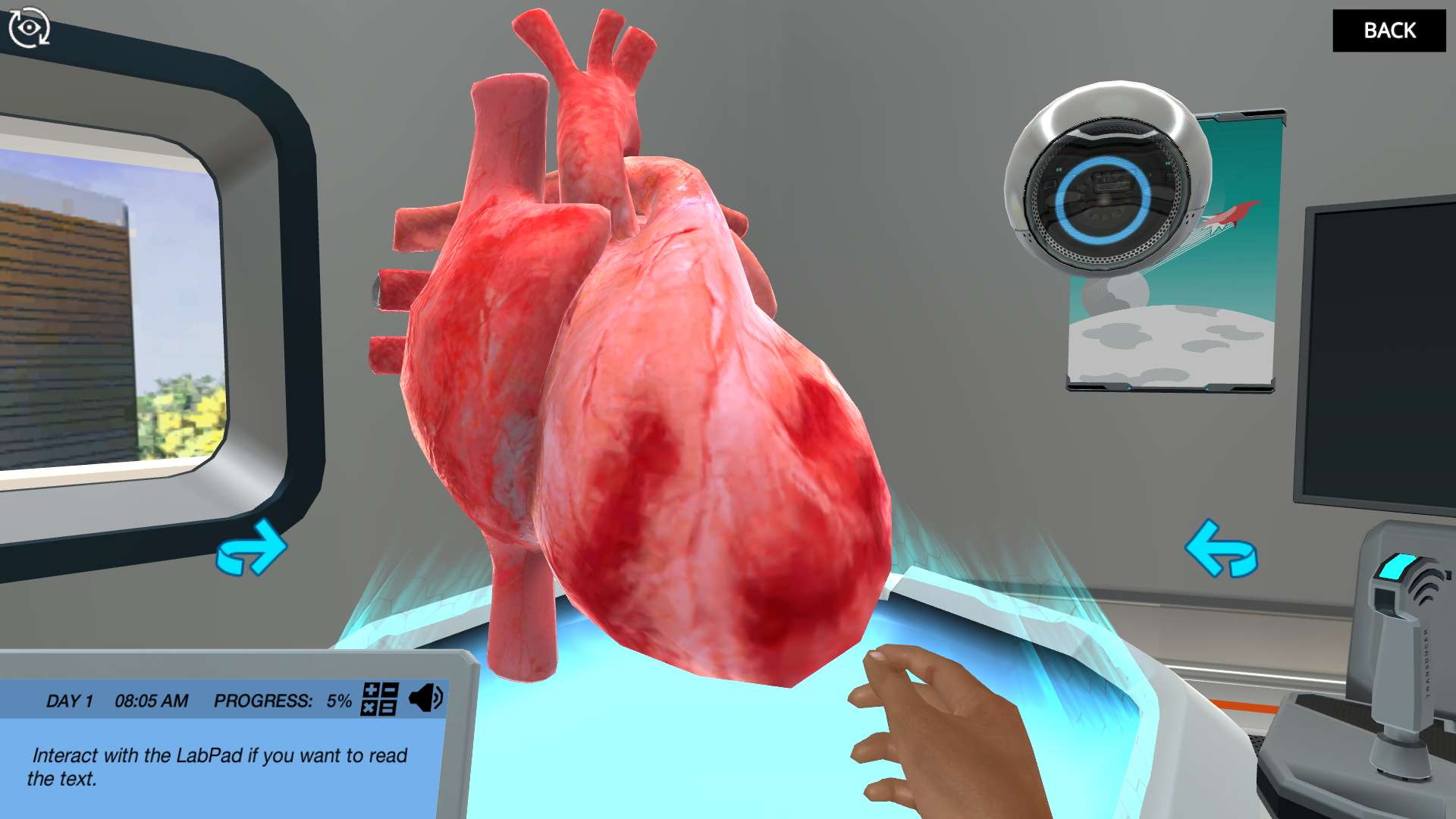
Practice obtaining different ultrasound views of the heart anatomy and use different Doppler ultrasound imaging techniques on a giant virtual heart. Apply your new skills to diagnose a patient.
Learning Objectives
- Distinguish and apply the different projections used in a basic echocardiography examination, as well as where the transducer is placed to obtain them.
- Relate the position and angle of the transducer as well as direction of its indicator to certain projections.
- Identify anatomical landmarks in the different projections.
- Approach a patient with respect and confirm that it is the correct person (checking ID)
- Understand the physics behind Doppler and how and when to apply it correctly.
- Understand how and when to apply M-mode.
- Understand and evaluate the most common measurements used for evaluation of left and right ventricular systolic function.
- Understand and evaluate the most common measurements used for evaluation of left ventricular diastolic function.
- Assess heart chamber dimensions (left and right ventricles, left and right atria, aortic root, vena cava, valvular function) and recognize what makes a case normal.
About This Simulation
Lab Techniques
- M-mode
- Tissue Doppler
- Continuous-wave Doppler
- Pulsed-wave Doppler
- Echocardiography
- Color Doppler
Related Standards
- Biology D.4 The heart
Learn More About This Simulation
Welcome to the Labster cardiology training facility! In this simulation, you will learn how to perform cardiac ultrasound using different modes and views. After practicing on a virtual heart, you will also evaluate cardiac function on a patient.
Use a giant virtual heart to practice Doppler ultrasound imaging
In our virtual training facility, we have a giant heart to practice the many different ultrasound transducer positions and angles that need to be mastered to correctly visualize and identify cardiac structures. It takes a lot of practice and sometimes small differences can make a difference when it comes to diagnosing abnormalities! In this simulation, you can repeat this training as much as you would like to familiarize yourself with normal cardiac structures, where the transducer needs to be placed, and at what angle to obtain the desired views.
Perform cardiac ultrasound on a patient
After your practice, you can choose to dust off your knowledge of ultrasound physics to better understand the technique and its different modes. Alternatively, you can also skip this lesson and immediately proceed to meet your patient, Mr. Johansson, a 50-year-old male who was referred here for an echocardiography examination. He presents symptoms of previous pain in the left arm and possibly in the chest, which he thought had to do with bad ergonomy at office work, being left-handed and working a lot on a computer. However, a physical exam from his physician revealed a systolic murmur in the stethoscope.
To get to the bottom of Mr. Johansson’s case and help inform his doctor, you will have to obtain the different ultrasound projections you practiced, as well as make some measurements and observations. Dr. One will be there to help you out, save your images and make notes of all the data.
Analyze the data and diagnose the patient
At the end of this very thorough examination, you will have access to all the images, videos and measurements that you performed on the patient, as well as on the practice healthy heart. By understanding and comparing all the information you have now gathered, will you be able to correctly diagnose the patient?
For Science Programs Providing a Learning Advantage
Boost STEM Pass Rates
Boost Learning with Fun
75% of students show high engagement and improved grades with Labster
Discover Simulations That Match Your Syllabus
Easily bolster your learning objectives with relevant, interactive content
Place Students in the Shoes of Real Scientists
Practice a lab procedure or visualize theory through narrative-driven scenarios


FAQs
Find answers to frequently asked questions.
Heading 1
Heading 2
Heading 3
Heading 4
Heading 5
Heading 6
Lorem ipsum dolor sit amet, consectetur adipiscing elit, sed do eiusmod tempor incididunt ut labore et dolore magna aliqua. Ut enim ad minim veniam, quis nostrud exercitation ullamco laboris nisi ut aliquip ex ea commodo consequat. Duis aute irure dolor in reprehenderit in voluptate velit esse cillum dolore eu fugiat nulla pariatur.
Block quote
Ordered list
- Item 1
- Item 2
- Item 3
Unordered list
- Item A
- Item B
- Item C
Bold text
Emphasis
Superscript
Subscript
A Labster virtual lab is an interactive, multimedia assignment that students access right from their computers. Many Labster virtual labs prepare students for success in college by introducing foundational knowledge using multimedia visualizations that make it easier to understand complex concepts. Other Labster virtual labs prepare learners for careers in STEM labs by giving them realistic practice on lab techniques and procedures.
Labster’s virtual lab simulations are created by scientists and designed to maximize engagement and interactivity. Unlike watching a video or reading a textbook, Labster virtual labs are interactive. To make progress, students must think critically and solve a real-world problem. We believe that learning by doing makes STEM stick.
Yes, Labster is compatible with all major LMS (Learning Management Systems) including Blackboard, Canvas, D2L, Moodle, and many others. Students can access Labster like any other assignment. If your institution does not choose an LMS integration, students will log into Labster’s Course Manager once they have an account created. Your institution will decide which is the best access method.
Labster is available for purchase by instructors, faculty, and administrators at education institutions. Purchasing our starter package, Labster Explorer, can be done using a credit card if you are located in the USA, Canada, or Mexico. If you are outside of North America or are choosing a higher plan, please speak with a Labster sales representative. Compare plans.
Labster supports a wide range of STEM courses at the high school, college, and university level across fields in biology, chemistry, physics, and health sciences. You can identify topics for your courses by searching our Content Catalog.


.JPG?fm=jpg&w=450&h=400)


