Heading 1
Heading 2
Heading 3
Heading 4
Heading 5
Heading 6
Lorem ipsum dolor sit amet, consectetur adipiscing elit, sed do eiusmod tempor incididunt ut labore et dolore magna aliqua. Ut enim ad minim veniam, quis nostrud exercitation ullamco laboris nisi ut aliquip ex ea commodo consequat. Duis aute irure dolor in reprehenderit in voluptate velit esse cillum dolore eu fugiat nulla pariatur.
Block quote
Ordered list
- Item 1
- Item 2
- Item 3
Unordered list
- Item A
- Item B
- Item C
Bold text
Emphasis
Superscript
Subscript
About This Simulation
Analyze the microscopic structure of the small intestine and learn the advantages and limitations of light, fluorescence and electron microscopy.
Learning Objectives
- Understand different microscopy techniques and their limitations
- Identify various cell types and cellular structures
- Understand coeliac disease and intestinal inflammation
- Understand staining techniques
About This Simulation
Lab Techniques
- Fluorescence Microscopy
- Light microscopy: using immersion oil and working with different objectives
Related Standards
- Not directly assessed, could support many areas depending on educators approach
- Biology Unit 2: Cell Structure and Function
- 1.1 Introduction to cells*
- 1.2 Ultrastructure of cells
Learn More About This Simulation
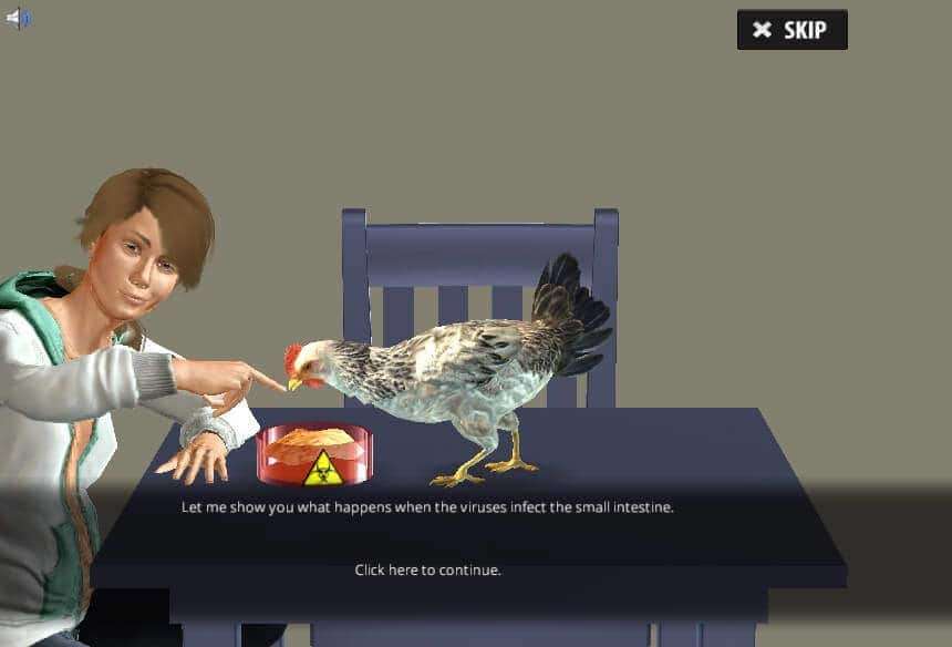
Join the Microscopy lab and learn about the different types of microscopy to understand the mechanisms behind it. You will be trained in light microscopy, transmission electron microscopy and fluorescence microscopy.
Use magnification
In the Microscopy lab, you will be presented with chicken intestinal slides that have been stained with Anilin, Orange G and Fuchsin. Using the 5x magnification, you will identify the villus and then proceed with higher magnifications to identify smooth muscle, Relatedcellular tissue, epithelial cells, Goblet cells and the nuclei.
Try out the electron microscope
Electron microscopes can be used to visualize objects that are too small to see when using a light microscope—for example, the microvilli, mitochondria and the junctions between cells. In the Microscopy lab, you will examine a chicken intestine slide that is specially prepared for a transmission electron microscope. You can zoom in and out to observe different cellular structures.
Learn about fluorescence staining techniques
You will learn about fluorescence staining techniques and how they can be used to visualize specific structures. For example, by staining the DNA with DAPI, you can easily identify a cell’s nucleus. In this part of the lab, you will examine a chicken intestine sample that is infected with a retrovirus and observe how the virus infects the lymphocytes and how it inhibits inflammation. The retrovirus can be further developed as a medicine for coeliac disease.
For Science Programs Providing a Learning Advantage
Boost STEM Pass Rates
Boost Learning with Fun
75% of students show high engagement and improved grades with Labster
Discover Simulations That Match Your Syllabus
Easily bolster your learning objectives with relevant, interactive content
Place Students in the Shoes of Real Scientists
Practice a lab procedure or visualize theory through narrative-driven scenarios


FAQs
Find answers to frequently asked questions.
Heading 1
Heading 2
Heading 3
Heading 4
Heading 5
Heading 6
Lorem ipsum dolor sit amet, consectetur adipiscing elit, sed do eiusmod tempor incididunt ut labore et dolore magna aliqua. Ut enim ad minim veniam, quis nostrud exercitation ullamco laboris nisi ut aliquip ex ea commodo consequat. Duis aute irure dolor in reprehenderit in voluptate velit esse cillum dolore eu fugiat nulla pariatur.
Block quote
Ordered list
- Item 1
- Item 2
- Item 3
Unordered list
- Item A
- Item B
- Item C
Bold text
Emphasis
Superscript
Subscript
A Labster virtual lab is an interactive, multimedia assignment that students access right from their computers. Many Labster virtual labs prepare students for success in college by introducing foundational knowledge using multimedia visualizations that make it easier to understand complex concepts. Other Labster virtual labs prepare learners for careers in STEM labs by giving them realistic practice on lab techniques and procedures.
Labster’s virtual lab simulations are created by scientists and designed to maximize engagement and interactivity. Unlike watching a video or reading a textbook, Labster virtual labs are interactive. To make progress, students must think critically and solve a real-world problem. We believe that learning by doing makes STEM stick.
Yes, Labster is compatible with all major LMS (Learning Management Systems) including Blackboard, Canvas, D2L, Moodle, and many others. Students can access Labster like any other assignment. If your institution does not choose an LMS integration, students will log into Labster’s Course Manager once they have an account created. Your institution will decide which is the best access method.
Labster is available for purchase by instructors, faculty, and administrators at education institutions. Purchasing our starter package, Labster Explorer, can be done using a credit card if you are located in the USA, Canada, or Mexico. If you are outside of North America or are choosing a higher plan, please speak with a Labster sales representative. Compare plans.
Labster supports a wide range of STEM courses at the high school, college, and university level across fields in biology, chemistry, physics, and health sciences. You can identify topics for your courses by searching our Content Catalog.


