Heading 1
Heading 2
Heading 3
Heading 4
Heading 5
Heading 6
Lorem ipsum dolor sit amet, consectetur adipiscing elit, sed do eiusmod tempor incididunt ut labore et dolore magna aliqua. Ut enim ad minim veniam, quis nostrud exercitation ullamco laboris nisi ut aliquip ex ea commodo consequat. Duis aute irure dolor in reprehenderit in voluptate velit esse cillum dolore eu fugiat nulla pariatur.
Block quote
Ordered list
- Item 1
- Item 2
- Item 3
Unordered list
- Item A
- Item B
- Item C
Bold text
Emphasis
Superscript
Subscript
About This Simulation
Join this virtual confocal microscopy lab and learn how to take pin-sharp confocal micrographs and 3D renderings. Use the knowledge to save your uncle’s crop from a mysterious plant disease.
Learning Objectives
- Describe the principles of confocal microscopy
- Use the basic functions of a confocal microscope
- Select the optimal settings to take confocal micrographs
- Acquire confocal images and create 3D renderings
- Describe the setup of a confocal microscope
- Discuss the advantages of confocal microscopy over conventional optical microscopy
About This Simulation
Lab Techniques
- Laser scanning confocal microscopy
- Staining of plant samples for fluorescence microscopy
Related Standards
- Early Stage Bachelors Level
- Early Stage Masters Level
- EHEA First Cycle
- EHEA Second Cycle
- FHEQ 6
- FHEQ 7
- Intermediate Stage Bachelors Level
- Late Stage Bachelors Level
- US College Year 1
- US College Year 2
- US College Year 3
- US College Year 5
- US College Year 4
- Support for laboratory skills at all levels
Learn More About This Simulation
Are you tired of blurry fluorescent microscope images and do you want to move to the next level? In the Confocal Microscopy simulation, you will learn about the benefits of using a confocal microscope over other optical imaging techniques. You’ll get to take a closer look at the setup of this type of microscope and learn how to operate it.
Identify the plant disease
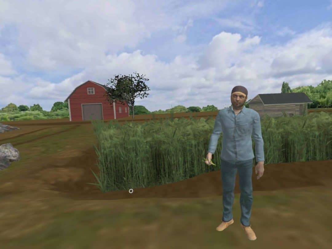
Your overall task will be to investigate a mysterious plant disease found at your uncle’s farm. You’ll make a visit to the farm and take an infected barley sample back to the lab where you canl take a closer look at the specimen by using confocal microscopy.
Take a look inside the microscope
In the Confocal Microscopy lab, you’ll be able to look inside a confocal microscope. Follow the lightpath along the mirrors and through the pinhole, and you’ll soon learn how the powerful light of a laser is used to scan a sample of a single layer. See how the pinhole contributes to the formation of razor-sharp fluorescent images.
Analyze the 3D structure
Your last task in the Confocal Microscopy lab is to choose the optimal exposure and scan line speed to take the best possible fluorescence images of the stained crop sample. Finally, you will take a stack of confocal images to create a 3D rendering that will help you to investigate what has infected your uncle’s barley.
Are you ready to identify the plant disease and give your uncle advice on a potential treatment to save his crops
For Science Programs Providing a Learning Advantage
Boost STEM Pass Rates
Boost Learning with Fun
75% of students show high engagement and improved grades with Labster
Discover Simulations That Match Your Syllabus
Easily bolster your learning objectives with relevant, interactive content
Place Students in the Shoes of Real Scientists
Practice a lab procedure or visualize theory through narrative-driven scenarios


FAQs
Find answers to frequently asked questions.
Heading 1
Heading 2
Heading 3
Heading 4
Heading 5
Heading 6
Lorem ipsum dolor sit amet, consectetur adipiscing elit, sed do eiusmod tempor incididunt ut labore et dolore magna aliqua. Ut enim ad minim veniam, quis nostrud exercitation ullamco laboris nisi ut aliquip ex ea commodo consequat. Duis aute irure dolor in reprehenderit in voluptate velit esse cillum dolore eu fugiat nulla pariatur.
Block quote
Ordered list
- Item 1
- Item 2
- Item 3
Unordered list
- Item A
- Item B
- Item C
Bold text
Emphasis
Superscript
Subscript
A Labster virtual lab is an interactive, multimedia assignment that students access right from their computers. Many Labster virtual labs prepare students for success in college by introducing foundational knowledge using multimedia visualizations that make it easier to understand complex concepts. Other Labster virtual labs prepare learners for careers in STEM labs by giving them realistic practice on lab techniques and procedures.
Labster’s virtual lab simulations are created by scientists and designed to maximize engagement and interactivity. Unlike watching a video or reading a textbook, Labster virtual labs are interactive. To make progress, students must think critically and solve a real-world problem. We believe that learning by doing makes STEM stick.
Yes, Labster is compatible with all major LMS (Learning Management Systems) including Blackboard, Canvas, D2L, Moodle, and many others. Students can access Labster like any other assignment. If your institution does not choose an LMS integration, students will log into Labster’s Course Manager once they have an account created. Your institution will decide which is the best access method.
Labster is available for purchase by instructors, faculty, and administrators at educational institutions. You can purchase Labster by speaking with a Labster sales representative. Compare plans.
Labster supports a wide range of STEM courses at the high school, college, and university level across fields in biology, chemistry, physics, and health sciences. You can identify topics for your courses by searching our Content Catalog.


.png?fm=jpg&w=450&h=400)


